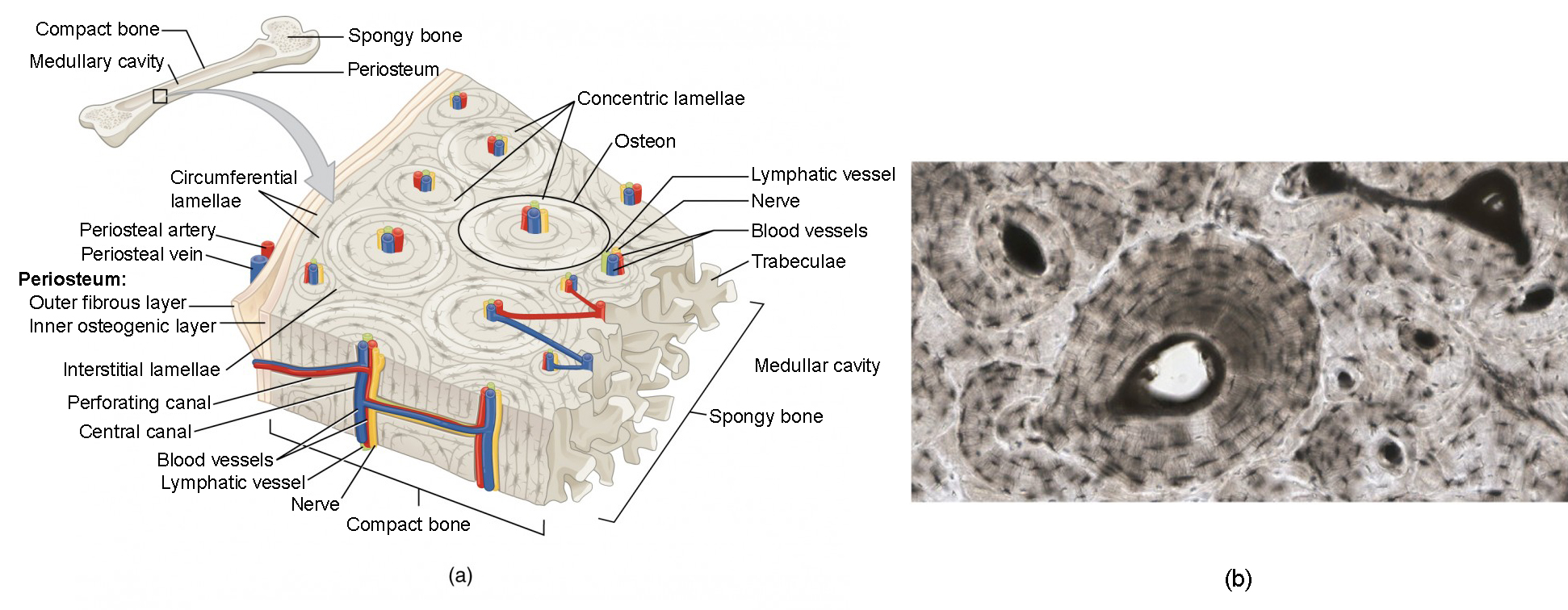
Cross Section Of A Bone / What are antlers and why do deer have them? | Wildlife Online / Cross section of a bone :. Compact bone, spongy bone, and bone marrow. Why is the marrow red? Cross section of the shaft (diaphysis). The large dark spots are passages for blood vessels and nerves. An outer 'fibrous layer' containing mainly fibroblasts, and an inner 'cambium layer' containing progenitor cells.
There are trabeculae in spongy bone which gives its sponge like appearance. The large dark spots are passages for blood vessels and nerves. Cross section of a bone. This is a cross section through decalcified bone. Can you identify the concentric lamellae, central canal and the lacunae.
Cross Section Of A Bone : Dinosaur Bone Cross Section ... from s3-us-west-2.amazonaws.com I don't find it enhances the image. This photo shows a cross section through bone. Two types of bone tissues in cross section of a long bone : Table 1 describes the bone markings, which are illustrated in (figure 4). Two types of bone tissues in cross section of a long bone : Smartdraw includes 1000s of professional healthcare and anatomy chart templates that you can modify and make your own. Would it be a good thing to show the epiphyseal plate? Cross section of mandible at first molar region showing cortical and spongy bone basic concepts in osteogenesis bone is a dynamic biological tissue, composed of various metabolically active cells that are integrated into a rigid framework.
Compact bone cross section courtesy:
Compact bone is the outer layer and the spongy bone forms the inner layer. Now that you know what bones do, let's take a look at what they're made of and their anatomy. Decalcified compact bone looks completely different than compact bone that still has calcium salts in its matrix. To look at a cross section, you will need to find a bone that's broken or cut one to look inside it. Would it be a good thing to show the epiphyseal plate? Each bone in your body is made up of three main types of bone material: Compact bone cross section courtesy: As the names suggest compact bone looks compact and the spongy bone looks like sponges. They are obtained by taking imaginary slices perpendicular to the main axis of organs, vessels, nerves, bones, soft tissue, or even the entire human body. Compact bone, spongy bone, and bone marrow. This is known as the periosteum. Find the perfect bone cross section stock photos and editorial news pictures from getty images. Two types of bone tissues in cross section of a long bone :
This is a short tutorial using blender 2.8 that shows how to create a bone cross section and. If i can teach you one thing about how to draw the back of a person, it's that it's absolutely crucial to understand the position of the scapula bones (shoulder in this tutorial, we will go over the bones and major muscle groups you will need to know to draw the. Cross section of a human bone showing bone marrow, spongy bone and blood vessels. This is a cross section through decalcified bone. The cross section of a rectangular pyramid is a rectangle.
5- Bone - Histology with Garland at University of ... from classconnection.s3.amazonaws.com An outer 'fibrous layer' containing mainly fibroblasts, and an inner 'cambium layer' containing progenitor cells. Bone markings the surface features of bones vary considerably, depending on the function and location in the body. The central tubular region of the bone, called the diaphysis, flares outward near the end to form the metaphysis, which contains a largely cancellous, or spongy, interior. Compact bone cross section courtesy: Bone matrix and cells bone matrix osseous tissue is a connective tissue and like all connective tissues contains relatively few cells and large amounts of extracellular matrix. Each bone in your body is made up of three main types of bone material: The surface features of bones vary considerably, depending on the function and location in the body. Related posts of cross section of human bone diagram human back muscles and bones.
Y points to the middle layer.
And why does the marrow stop where it does, and so sharply? Histology slide courtesy of william l. A cross sectional view of bone. Human back muscles and bones 12 photos of the human back muscles and bones human back muscles and bones, bone, human back muscles and bones Would it be a good thing to show the epiphyseal plate? Smartdraw includes 1000s of professional healthcare and anatomy chart templates that you can modify and make your own. The head of each end of a long bone consists largely of spongy bone and is covered with hyaline cartilage. Two types of bone tissues in cross section of a long bone : Human bone, cross section diagram of femur showing osteon, veins, marrow. Browse 53 bone marrow cross section stock photos and images available, or search for bone cross section or bone cells to find more great stock photos and pictures. Bone and bones / pathology*. A long bone illustrates both types of bone. Table 1 describes the bone markings, which are illustrated in (figure 4).
Which structures of the bone are indicated by the arrows? The cross section of a rectangular pyramid is a rectangle. Online quiz to learn structure of a long bone (humerus). They are obtained by taking imaginary slices perpendicular to the main axis of organs, vessels, nerves, bones, soft tissue, or even the entire human body. Find the perfect bone cross section stock photos and editorial news pictures from getty images.
Cross section through an model of an normal upper right ... from c8.alamy.com And why does the marrow stop where it does, and so sharply? I don't find it enhances the image. A long bone illustrates both types of bone. Table 1 describes the bone markings, which are illustrated in (figure 4). Marrow in the shaft of long bones is typically yellow, with red marrow in the head through the cancellous bone. The central tubular region of the bone, called the diaphysis, flares outward near the end to form the metaphysis, which contains a largely cancellous, or spongy, interior. Internal structure of a human long bone internal structure of a human long bone, with a magnified cross section of the interior. This photo shows a cross section through bone.
Would it be a good thing to show the epiphyseal plate?
Cross section of mandible at first molar region showing cortical and spongy bone basic concepts in osteogenesis bone is a dynamic biological tissue, composed of various metabolically active cells that are integrated into a rigid framework. The surface features of bones vary considerably, depending on the function and location in the body. The central tubular region of the bone, called the diaphysis, flares outward near the end to form the metaphysis, which contains a largely cancellous, or spongy, interior. This is a cross section through decalcified bone. Compact bone cross section courtesy: Bone markings the surface features of bones vary considerably, depending on the function and location in the body. Why is the marrow red? The humerus is the long bone located in the upper arm of the body which extends from the shoulder joint to the elbow. X points to the inner layer. The compact bone is made up of osteon. A cross sectional view of bone. This is a short tutorial using blender 2.8 that shows how to create a bone cross section and. Browse 53 bone marrow cross section stock photos and images available, or search for bone cross section or bone cells to find more great stock photos and pictures.